By IDSE News Staff
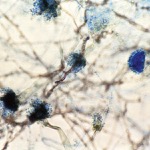
A new imaging method will allow specific detection of Aspergillus fumigatus fungal infections faster, without the need for invasive procedures. Delays in diagnosing fungal infections caused by Aspergillus and other fungi can put immunocompromised patients at risk for more serious illnesses or even death, said researchers from the National Institutes of Health.
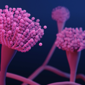
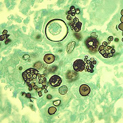
Due to their presence in the environment, many fungi evolved to use other sources of fuel besides glucose, such as by breaking down complex sugars into simple ones to produce energy. Aspergillus can break down a specific sugar, cellobiose, into two glucose molecules, while most other microbes and human cells cannot. The researchers developed a radioactive version of cellobiose, which when injected in the blood, it can be visualized in the body using PET scanners.
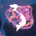
In this study, radioactive cellobiose ([18F]-fluorocellobiose, [18F]-FCB) was injected in mice with fungal infections that were then imaged using a specialized PET scanner for small animals. The mice showed accumulation of radioactivity, while mice with bacterial infections or noninfectious inflammation did not (Sci Transl Med 14 Aug 2024. doi:10.1126/scitranslmed.adl5934).
Researchers also found that the same radioactive tracer, [18F]-FCB, can determine whether the mice with fungal infections are responding to treatment through PET images taken before and after starting treatment.
The study was funded by the Center for Infectious Disease Imaging, a joint initiative between Radiology and Imaging Sciences at the NIH Clinical Center and the National Institute of Allergy and Infectious Diseases, in collaboration with the Chemistry and Synthesis Center at the National Heart, Lung, and Blood Institute.